
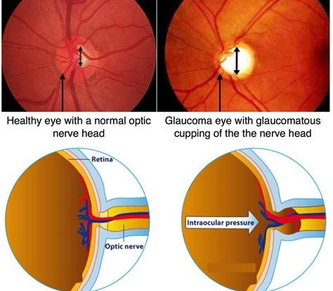
Intraocular pressure (IOP) is the fluid pressure inside the eye. High IOP can lead to glaucoma, a serious eye disease that can cause vision loss.
Intraocular pressure is a term that might not be familiar to many, but it’s a condition that affects millions of people worldwide. With the potential to lead to blindness if left untreated, it’s a serious issue that demands attention. But what exactly is intraocular pressure, and why is it so concerning? To understand this condition, we need to explore the anatomy of the eye and how it functions.
Firstly, the eye is a complex organ that requires a delicate balance of pressure to function correctly. Intraocular pressure refers to the fluid pressure inside the eye that helps maintain its shape and nourish the tissues. However, when this pressure becomes too high or too low, it can cause significant damage to the optic nerve and lead to vision loss. This is why monitoring intraocular pressure is critical for those at risk of developing glaucoma, a condition that affects over 60 million people globally.
So, what are the risk factors for developing high intraocular pressure, and how can it be managed? These are questions that ophthalmologists and researchers have been exploring for decades, with exciting advances in technology and treatment options emerging in recent years. In this article, we’ll delve deeper into the world of intraocular pressure and explore what it means for your eye health.
Daftar Isi
The Importance of Intraocular Pressure
When it comes to our eyesight, there is one crucial factor that we should always keep in mind: intraocular pressure. This refers to the pressure inside the eye, specifically within the anterior chamber where the aqueous humor flows. Intraocular pressure helps maintain the shape of the eye and provides nutrients to the tissues, but when it becomes too high or too low, it can lead to serious vision problems.
How Intraocular Pressure is Measured
Measuring intraocular pressure is a standard part of a comprehensive eye exam. It is usually done using a tonometer, which can either be a handheld device that lightly touches the cornea or a machine that releases a puff of air onto the eye. Normal intraocular pressure ranges between 10-21 mmHg, but this can vary depending on factors such as age and medical history.
Glaucoma and High Intraocular Pressure
One of the most significant risks associated with high intraocular pressure is glaucoma. This condition damages the optic nerve and can lead to permanent vision loss if left untreated. Glaucoma is often called the silent thief of sight because it typically has no symptoms until significant damage has occurred. People over the age of 60, those with a family history of glaucoma, and individuals with certain medical conditions are at higher risk of developing glaucoma.
Treatment Options for High Intraocular Pressure
If diagnosed with high intraocular pressure, there are several treatment options available. Eye drops are often the first line of defense, as they can help reduce intraocular pressure by either decreasing the amount of fluid produced in the eye or increasing its outflow. Other treatments include laser therapy, which can help open up the eye’s drainage system, and surgery, which can create a new drainage channel in the eye.
Hypotony and Low Intraocular Pressure
While high intraocular pressure is often associated with glaucoma, low intraocular pressure (hypotony) can also cause vision problems. Hypotony occurs when the pressure inside the eye drops below normal levels, usually due to surgery or trauma. Symptoms of hypotony can include blurry vision, double vision, and eye pain.
Treatment Options for Low Intraocular Pressure
The treatment for hypotony will depend on the underlying cause. If it is due to surgery, the surgeon may need to perform additional procedures to correct the issue. If it is due to trauma, the eye may need to be monitored closely to ensure that the pressure returns to normal levels on its own. In some cases, a temporary patch or bandage contact lens may be used to help the eye heal.
The Importance of Regular Eye Exams
Whether you are at risk for high or low intraocular pressure, it is vital to schedule regular eye exams. During these exams, your eye doctor can check your intraocular pressure, evaluate your overall eye health, and detect any potential issues early on. By catching problems early, you can receive prompt treatment and protect your vision for the long term.
Preventing High Intraocular Pressure
While some risk factors for high intraocular pressure, such as age and family history, cannot be changed, there are steps you can take to protect your eyesight. Maintaining a healthy weight, eating a balanced diet, and exercising regularly can all help reduce your risk of developing high intraocular pressure. Additionally, avoiding smoking and limiting your alcohol intake can also improve your eye health.
The Bottom Line on Intraocular Pressure
When it comes to your vision, intraocular pressure is a critical factor that should not be overlooked. Regular eye exams, healthy lifestyle habits, and prompt treatment when necessary can all help protect your eyesight and prevent serious vision problems. By being proactive about your eye health, you can enjoy clear vision for years to come.
What is Intraocular Pressure and Why is it Important?
Intraocular pressure (IOP) refers to the pressure inside the eye. It is a vital measurement in assessing the health of the eye and diagnosing conditions such as glaucoma. The eye needs to maintain a delicate balance of fluid production and drainage to maintain its shape and function properly. IOP is an indication of this balance and if it becomes too high, it can cause damage to the optic nerve and lead to vision loss.
The Anatomy of the Eye: Understanding Intraocular Pressure
The eye is filled with a clear, gel-like substance called the vitreous humor. Behind the iris and pupil lies the lens, which focuses incoming light onto the retina at the back of the eye. The retina contains photoreceptor cells that convert light into electrical signals that travel through the optic nerve to the brain for interpretation. The ciliary body, located behind the iris, produces the aqueous humor, a clear fluid that fills the space between the cornea and the iris. This fluid provides nutrients to the surrounding tissues and helps maintain the eye’s shape. Excess aqueous humor drains out of the eye through a network of channels called the trabecular meshwork, located at the angle where the iris meets the cornea. This drainage system needs to function correctly to maintain a healthy IOP.
How is Intraocular Pressure Measured?
IOP is measured using a tonometer, which measures the force needed to flatten the cornea. There are several methods of tonometry, including applanation tonometry, where a small amount of pressure is applied to the cornea while the patient looks at a target. Non-contact tonometry uses a puff of air directed at the cornea to measure the IOP. A normal IOP range is typically between 10-21 mmHg (millimeters of mercury). However, this range can vary depending on age, race, and other factors. It is important to note that a single IOP reading does not provide an accurate assessment of eye health, and multiple readings over time may be necessary for diagnosis.
Glaucoma: When Intraocular Pressure Becomes a Problem
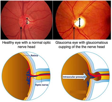
Glaucoma is a group of eye conditions that damage the optic nerve and can lead to vision loss. It is often associated with high IOP, although some people with glaucoma have normal IOP levels. There are two main types of glaucoma: open-angle and angle-closure. Open-angle glaucoma is the most common type and develops slowly over time. Angle-closure glaucoma is less common but can develop rapidly and cause severe symptoms such as eye pain, nausea, and blurred vision. Glaucoma is a leading cause of blindness worldwide, and early detection and treatment are crucial in preventing vision loss.
Risk Factors for High Intraocular Pressure and Glaucoma
Age, family history, and certain medical conditions such as diabetes and high blood pressure can increase the risk of developing high IOP and glaucoma. Other risk factors include nearsightedness, a history of eye injuries or surgeries, and the use of corticosteroid medications. People of African descent are also at higher risk for developing glaucoma. Regular eye exams and screening for glaucoma are recommended for people at higher risk.
Symptoms and Effects of High Intraocular Pressure
High IOP does not typically cause noticeable symptoms until it has reached a severe level. Symptoms that may indicate high IOP include blurred vision, halos around lights, headache, and eye pain. However, these symptoms can also be caused by other conditions, so it is important to have a comprehensive eye exam to determine the cause. If left untreated, high IOP can cause damage to the optic nerve and lead to glaucoma and vision loss.
Treatment Options for High Intraocular Pressure and Glaucoma
The goal of treatment for high IOP and glaucoma is to lower the pressure inside the eye to prevent further damage to the optic nerve. Treatment options may include eye drops, laser therapy, or surgery. Eye drops are typically the first line of treatment and work by either reducing the production of aqueous humor or increasing its drainage. Laser therapy can be used to open up the trabecular meshwork and improve drainage. Surgery may be necessary for people who do not respond to other treatments or whose glaucoma is severe. Treatment plans may vary depending on the type and severity of glaucoma and should be tailored to each individual’s needs.
New Developments in Intraocular Pressure Management
Recent advancements in technology have led to new methods of managing IOP and glaucoma. One such development is the use of micro-invasive glaucoma surgery (MIGS), which uses minimally invasive techniques to improve drainage and reduce IOP. Another promising area of research is the use of gene therapy to treat glaucoma by targeting specific genes involved in the disease process. These new developments offer hope for more effective and less invasive treatments for glaucoma in the future.
Lifestyle Modifications to Reduce Intraocular Pressure
While medical treatments are important in managing IOP and glaucoma, lifestyle modifications can also play a role in reducing IOP and improving overall eye health. Regular exercise, maintaining a healthy weight, and avoiding smoking can help reduce the risk of developing glaucoma. Some studies have also suggested that a diet rich in fruits and vegetables, particularly those high in antioxidants such as vitamins A, C, and E, may be beneficial for eye health.
The Importance of Regular Eye Exams for Monitoring Intraocular Pressure
Regular eye exams are an essential part of maintaining eye health and detecting conditions such as glaucoma. During an eye exam, your eye doctor will measure your IOP and assess your overall eye health. They may also perform additional tests such as visual field testing and optic nerve imaging to determine if there is any damage to the optic nerve. Early detection and treatment of high IOP and glaucoma are crucial in preventing vision loss and maintaining good eye health.
Intraocular pressure, also known as IOP, is a crucial measurement in ophthalmology that determines the pressure inside the eye. This pressure is vital as it can lead to various eye diseases such as glaucoma, which can cause permanent vision loss if not treated in time.Pros:1. Early detection of eye diseases: Regular measurement of IOP can help detect eye diseases such as glaucoma at an early stage, enabling prompt treatment and preventing vision loss.2. Easy to measure: Measuring IOP is a quick and non-invasive procedure that can be performed during a routine eye exam.3. Helps monitor treatment progress: For patients with eye diseases such as glaucoma, regular IOP measurement can help monitor the effectiveness of treatment and adjust it accordingly.Cons:1. Inaccurate readings: IOP measurements can sometimes be inaccurate due to various factors such as corneal thickness, patient position, or use of certain medications.2. False positives: High IOP readings do not always indicate glaucoma, leading to unnecessary anxiety and further testing for patients.3. Patient discomfort: Some patients may experience discomfort or mild pain during the IOP measurement procedure.In conclusion, while IOP measurement is an essential tool in identifying and managing eye diseases, its limitations should also be considered. It is important for ophthalmologists to educate their patients on the importance of regular IOP measurement and to interpret the results accurately to ensure the best possible outcomes for their patients’ eye health.
As a journalist, it is my responsibility to inform you about the importance of intraocular pressure. Intraocular pressure refers to the pressure within the eye, which is crucial in maintaining the shape and function of the eye. It is essential to have a balanced intraocular pressure to prevent any damage to the optic nerve and avoid vision loss.
High intraocular pressure is a common symptom of glaucoma, a condition that can cause permanent vision loss if left untreated. It is vital to schedule regular eye exams to detect any changes in intraocular pressure and receive prompt treatment if necessary. Treatment options for high intraocular pressure may include medication or surgery, depending on the severity of the condition.
In conclusion, intraocular pressure is an important aspect of your eye health that should not be overlooked. By staying informed and seeking regular eye exams, you can take measures to maintain healthy intraocular pressure and prevent any potential damage to your vision. Remember to prioritize your eye health and schedule an appointment with an eye specialist if you experience any changes in your vision or eye discomfort.
Video intraocular pressure
As a journalist, it is important to address the concerns of the public regarding their health. One common question that people ask is about intraocular pressure.
Here are some of the questions that people commonly ask about intraocular pressure:
- What is intraocular pressure?
- What is the normal range for intraocular pressure?
- Can high intraocular pressure cause vision loss?
- What are the symptoms of high intraocular pressure?
- How is intraocular pressure measured?
Let’s answer these questions one by one:
- What is intraocular pressure?
- What is the normal range for intraocular pressure?
- Can high intraocular pressure cause vision loss?
- What are the symptoms of high intraocular pressure?
- How is intraocular pressure measured?
Intraocular pressure is the pressure inside the eye. It is maintained by a balance between the production and drainage of aqueous humor, a clear fluid that fills the front part of the eye.
The normal range for intraocular pressure is between 12 and 22 mmHg (millimeters of mercury). However, some people may have higher or lower normal pressures.
Yes, high intraocular pressure can cause vision loss if it is not treated. It can lead to damage to the optic nerve, which is responsible for transmitting visual information from the eye to the brain.
Most people with high intraocular pressure do not have any symptoms. This is why regular eye exams are important to detect any changes in pressure or other eye conditions.
Intraocular pressure is measured using a device called a tonometer. There are two types of tonometers: the applanation tonometer and the non-contact tonometer. The applanation tonometer involves touching the cornea with a special probe, while the non-contact tonometer uses a puff of air to measure pressure.
It is important to remember that intraocular pressure is just one aspect of eye health. Regular eye exams are crucial to detect any changes in pressure and other eye conditions that may affect vision.